In a first, Indian scientists have revealed the microscopy images of SARS-CoV-2 virus or COVID-19 amid an ongoing battle against the coronavirus crisis. The scientists took the throat swab sample from first laboratory, the confirmed Coronavirus case in India, reported on January 30 in Kerala. The findings were published in the latest editon of the Indian Journal of Medical Research (IJMR).
HOW IJMR EXPLAINS THE MICROSCOPY IMAGES OF COVID-19
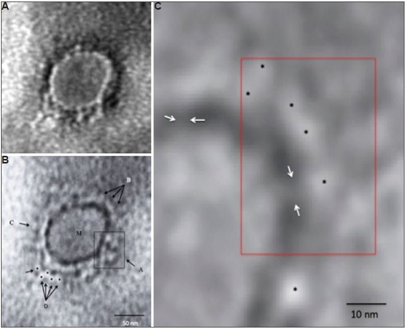
A. A representative negative-stained COVID-19 particle showing morphodiagnostic features of family Coronaviridae.
B. Defocussed image of the same particlle resolving the virus envelope glycoprotein morphology in finer details. The boxed area A shows a tetramer-like aggregate of four distinct peplomers, arrows shown by B show a more orthodox morphology of coronavirus surface projections. M indicates the matrix of the virus particle. C shows a distinct 'peplomer head' with negative stain silhouette. The area D is interesting as possible linear projections could be imaged. Five distinct peplomers could be imaged as shown by the arrows.
C. A highly magnified processed image for pixel corrections shows a distinct evidence of direct 'stalk' connecting the peplomer to the virion surface. The peplomers are shown with asterisk and the stalk with an arrow. Magnification bars are built into the micrographs.
ALSO READ | COVID-19 Crisis: 10-month-old infant tests positive for coronavirus in Karnataka