
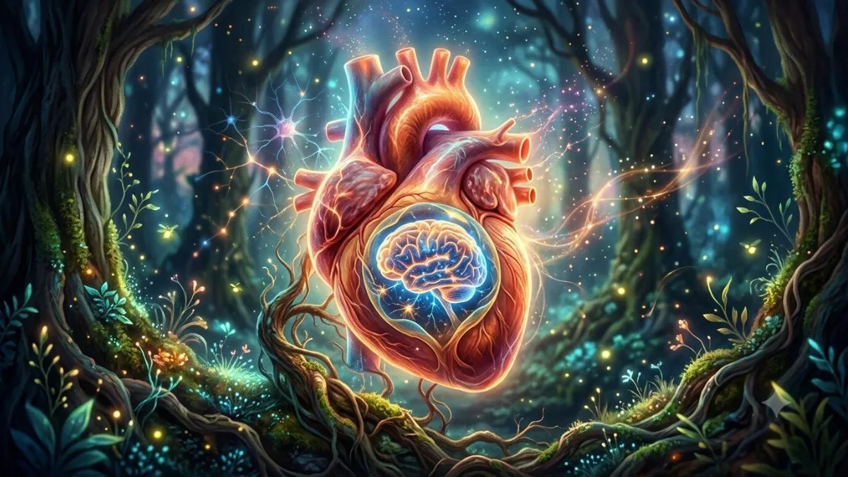
The heart has long been thought to follow instructions sent by the brain. But a new study suggests it may have a lot more independence than previously believed. Researchers have discovered a small network of neurons inside the heart that plays an active role in controlling heartbeats and protecting the organ during stressful situations.
The research, conducted at Yale University in the US and published in the scientific journal Cell in July 2026, found that this tiny nervous system helps regulate the heart's activity rather than simply passing on signals from the brain. The findings could improve scientists' understanding of how the heart functions and responds to physical and physiological stress.
Heart has its own 'brain'
The study focused on the intrinsic cardiac nervous system, a network of neurons found in the fatty tissue surrounding the heart. Although these nerve cells make up only around 0.01 per cent of the cells in that tissue, researchers found they act as a local control centre.
According to the research team, the neurons communicate with one another while also receiving signals from the brain, allowing them to take part in regulating the heart's activity.
How the heart's own nervous system works
The intestine is often referred to as the "second brain" because it contains a vast network of neurons involved in digestion and hormone production. The new study suggests the heart also has its own nervous system, although it is much smaller.
Researchers say this network forms the final stage of a complex neural circuit responsible for controlling cardiac activity.
To study it in detail, the team used genetically modified mice. They marked the neurons in the intrinsic cardiac nervous system and analysed them through genetic sequencing. This revealed two main groups of nerve cells, known as Npy+ neurons and Ddah1+ neurons.
The researchers found that each group performed a different role in how the heart functions and protects itself. Rather than simply relaying instructions from the brain, these neurons also process information locally and help determine how the heart should respond under different conditions.
The study noted that relatively little had been known about these cells or their importance in cardiovascular health. The new findings provide a clearer picture of how different neuron groups directly influence heartbeat regulation and heart protection.
Npy+ neurons help keep the heartbeat in check
The first set of experiments showed that Npy+ neurons are essential for regulating heart rate. When researchers stimulated these cells, the heart rate of the mice slowed down.
Removing the neurons had far more serious consequences. The animals developed heart failure and died after this group of cells was eliminated.
According to the study authors, the findings suggest that Npy+ neurons help slow the heart when it begins beating too quickly. They also appear to be essential for maintaining the heart's normal function.
Ddah1+ neurons become crucial when the heart is under stress
The second group of neurons behaved quite differently. During the initial experiments, activating or removing Ddah1+ neurons did not produce any significant changes in the animals. That initially led researchers to believe these cells had only a limited role.
However, an unexpected observation changed that view. During a routine blood pressure measurement, a student noticed that a mouse without these neurons died after the examination. The same outcome was later seen in other genetically modified animals.
That prompted the researchers to investigate whether Ddah1+ neurons were linked to stress responses. They exposed the mice to different forms of physical and physiological stress and found that most animals lacking these neurons were unable to cope with the conditions.
The findings suggest that while Ddah1+ neurons have little immediate effect on heartbeat under normal conditions, they become crucial when the body faces situations that require the heart to work harder. Without them, the heart appears to become far more vulnerable during periods of stress.
What the discovery could mean for human heart research
According to the researchers, the same neuron subtypes identified in mice were also found in humans. However, all of the functional experiments in this study were carried out only in animals.
Further research will be needed to determine whether Npy+ and Ddah1+ neurons perform the same roles in the human heart. If confirmed, the findings could deepen scientists' understanding of how the heart controls its rhythm and protects itself during stressful situations.
Overall, the study suggests the heart does more than simply follow instructions from the brain. It appears to have its own small neural network that can interpret signals and adjust heart activity according to the body's needs.
ALSO READ: